
The biggest map of the brain ever shows mouse neurons in amazing details
Neuroscientists have created the largest and detailed map of the mammals in a landmark achievement

Rendering of more than 1,000 brain cells from the mouse to be rebuilt from a cubic millimeter of brain.
Researchers have created the largest and most accurate Wiring diagram Current brain brain, mapping cells Cubic millimeter of a mouse brain tissue. In a landmark achievement, the diagram also determines the activity of individual neurons on a large scale and horbar; First Neuroscience.
The high-resolution 3D map has more than 200,000 brain cells, and about 82,000 of them are neurons. The synapse and more than 400 million neural cable over 4 kilometers. It is the only scale map to compare Cubic millimeter of human braiN, 16,000 neurons and 150 million signs of Sinapy. The new map captured thousands of thousands of neurons activity and interacts with each other to process visual information.
This brain activity map is combined with the wiring diagram, marks a landmark Connectomics aims to show how brains process and organize information. Behind massive efforts are more than 150 researchers in Inteligiruan Cortical Network (micro) projects, which worked in the eight papers currently published Nature and Nature methods. It has micro projects He made his resources available to the neuroscience community Online and other groups are already being studied in different studies.
To help Science Journalism
If you enjoy this article, consider entering award-winning journalism Subscribe. By purchasing subscription, you are helping to ensure the future of stories about the discoveries and ideas that are conformed to today.
“They managed to do something we have not done as a community in neuroscience in our entire history,” says Mariela Petkova, in the Neuroschusetts of the University of Cambridge, who do not participate in Massachusetts. “We have never seen this scale.”
The data is “very light,” says Forrest Collman, in Washington in Seattle, Washington, Washington, Washington, Neuroscientialist of the Brain Institute. “Looking at the gaze, it gives you a sense of complexity in the brain looking at the night stars.”
Mouse in a matrix
To create a map of progress, researchers fired the first 76,000 neurons while the cortex of a mouse saw several videos while watching various videos, including clips Matrixfor two hours. Then, a cubic millimeter of the mouse cut thousands of slices of thousands, each among the four hundred tissues of a human hair.
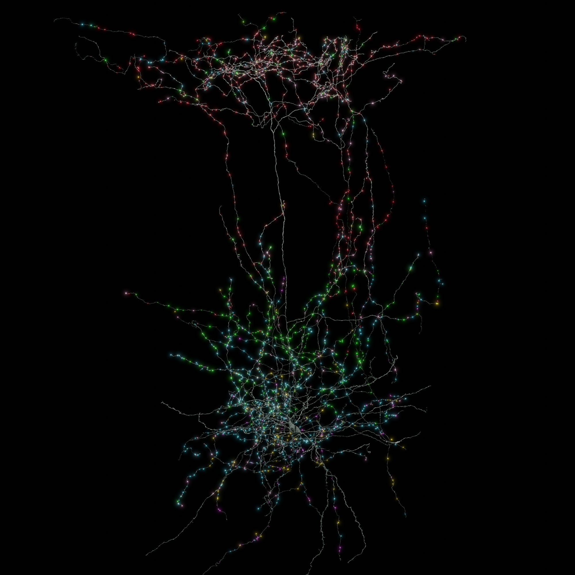
5. Layer Martinotti cell conversion (gray) Rebuilt from a large-scale electron set. Outgoing synapses (Shiny places) The colors of the target cell type are coded. Red dots are synapses of cold layers between 2/3 pyramidal cells and cyan points are Ixotous neurons in Telence TelenceFalic projects.
CLARRE GAMLIN / ALLEN Institute
Scientists represent each slice and images were mounted on the 3D map. Finally, artificial intelligence and machine learning algorithms used neurons to realize their branch projections and synapses. The team also has neurons on the map with the recording of brain cells.
Moritz Helmstaedter, Germany, the German Brain Research Max Planck Institute of Neuroscience, “combining function and structure scale” is unprecedented. “It’s a very impressive effort and success.”
Fire together, wire together
Work provided views on basic rules of form Nonural circuits Mouse in the brain. For example, the authors found neurons in the cortex that respond to similar visual features, such as movements or movement guidelines, often make up more connections with each other, whatever they do with neurons that specialize in other different characteristics.
The results add a new wrinkle to the long-time theory of neuroscience, Collman, namely “neurons that turn that wire”. Previous studies have tested this theory only in limited numbers of neurons and synapi. The current study shows that “there is also diversity (” that this rule applies to different components of Cortex, “he added.
Micro researchers hope their data set can help reveal various characteristics and processes in the brain. “There are coral sites we understand at different levels and different ways. And I think this is really the beginning of the structure and function,” says a author of the Author of the Clay Reide’s Institute and Microon.
Helmstaedter says that the researchers can use cable maps, how it stores brains and remembers visual memories, such as “Last birthday party or grandparents’ memories”. These are “open questions about very essential mami cortex,” he added.
The published map accounts for about 0.2% of the mouse brain, but technology will test the entire animal brain, says Nuno Maçarico is another author of the Neuroanatomista Institute and Micron.
This article reproduces with permission and has been First posted On April 9, 2025.